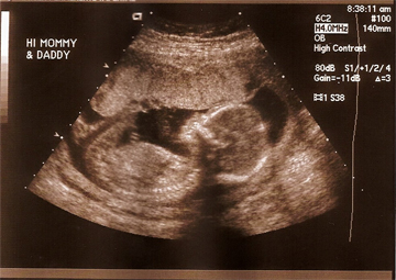
An ultrasound exam uses sound waves to scan a woman's abdomen and pelvic cavity, creating a picture (sonogram).
The sound waves bounce off tissues like echoes and are converted into images. Different substances, such as bones or fluid, change the sound waves so that they appear differently on the image. Liquid appears dark and dense tissue appears light or gray. This allows the ultrasound test to visualize bones, internal organs, amniotic fluid, or cysts.
Real-time ultrasound combines images into a recorded movie that can show movement. The movement of organs, such as the heart, also provides useful information.
There is no radiation involved in an ultrasound test.
Ultrasound during Pregnancy
Ultrasound tests are commonly ordered during pregnancy to evaluate the health of the fetus. Not all pregancies require a ultrasound examination.
The results of ultrasounds are usually evaluated along with the other prenatal tests, such as triple tests, amniocentesis, or chorionic villus sampling to provide a more complete understanding of the baby's health and potential problems.
An ultrasound exam is medically indicated throughout pregnancy for the following reasons:
- The age of the fetus. Ultrasound can provide a more accurate measure of the baby's age than the date of the last menstrual period (LMP). The crown-rump length, the distance from the crown of the head to the bottom, is one measure used to evaluate age.
- The rate of growth of the fetus. This is important to be sure that the baby is gaining appropriate weight for its age.
- Placement of the placenta. This is important to rule out placenta previa, the blockage of the birth canal by the placenta.
- Fetal heart rate and movement
- Fetal position. This is important to be sure that the baby is well-positioned for delivery and to rule out a breech presentation.
- Amount of amniotic fluid in the uterus. Too much or too little amniotic fluid suggests that there may be other medical problems to be evaluated.
- Multiples. Ultrasound can evaluate the presence of twins or triplets.
- Birth defects. Ultrasound can identify some, but not all, birth defects, such as spina bifida.
Ultrasound also may be used for diagnosing an ectopic pregnancy or determining a cause of bleeding or pain during pregnancy.
Ultrasound exams may be repeated during pregnancy if the woman has a high-risk pregnancy or the pregnancy continues after the due date.
Ultrasound for Women's Health
Ultrasound is also frequently performed to examine the pelvic organs in women who are not pregnant. It may be used for the following:
- Identify a pelvic mass
- Identify possible causes of pelvic pain
- Identify possible causes of abnormal bleeding or other menstrual problems
- Locate the position of an intrauterine device (IUD)
- Identify possible causes of infertility
Types of Ultrasound Exams
There are different types of ultrasound exams that may be ordered for different purposes.
- Transvaginal Scans. This ultrasound exam uses a narrow transducer (probe) that is inserted into the vagina to generate images. It can get closer to the uterus so it can provide clearer images of small masses. This is often used during the early stages of pregnancy.
- Standard Ultrasound. This is an ultrasound exam that uses a transducer over the abdomen. It creates 2-D images of the developing fetus .
- Advanced Ultrasound. This exam is similar to the standard ultrasound, uses more advanced equipment to provide better clarity of targeted organs that require additional evaluation.
- Doppler Ultrasound. Doppler can sense movement, such as blood flow, and can evaluate the heart and arteries.
- 3-D Ultrasound: This uses specially designed transducers and software to generate 3-D images of the developing fetus.
- 4-D or Dynamic 3-D Ultrasound: Uses specially designed scanners to look at the face and movements of the baby prior to delivery.
- Fetal Echocardiography: This is used to provide detailed images of the baby's heart and may be ordered to evaluate congenital heart disease.
What are the risks and side effects of ultrasound?
The ultrasound is a noninvasive procedure that, when used properly, has not demonstrated fetal harm. The long term effects of repeated ultrasound exposures on the fetus are not fully known. Ultrasound should only be used if medically indicated.
Frequently asked questions about ultrasound exams
If an ultrasound is done at 6 to 7 weeks and a heartbeat is not detected, does that mean there is a problem?
No it does not mean there is a problem. The heartbeat may not be detected that early for a variety of reasons, including inaccurate dating of the pregnancy. A Heartbeats are best detected with transvaginal ultrasounds early in pregnancy.
How accurate are ultrasounds in calculating gestational age?
Your healthcare provider will use hormone levels in your blood, the date of your last menstrual period and, in some cases, results from an ultrasound to generate an estimated gestational age. However, variations in each woman's cycle and each pregnancy may hinder the accuracy of the gestational age calculation. If your healthcare provider uses an ultrasound to get an estimated delivery date to base the timing of your prenatal care, the original estimated gestational age will not be changed.
When can an ultrasound determine whether the baby is a boy or a girl?
The fetus must usually have reached 18 to 20 weeks of gestation to determine its gender. The test is not 100% accurate in predicting whether they baby is a boy or girl. The position of the fetus and other factors will influence the accuracy of the test. To be absolutely sure....you must wait until the baby is born.
Are ultrasounds a necessary part of prenatal care?
Ultrasounds are only necessary if there is a medical concern. For women with an uncomplicated pregnancy, an ultrasound is not a necessary part of prenatal care.
Source: Office on Women's Health
Last updated : 1/8/2022